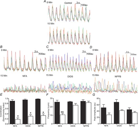
Figure 3. Loss of coordinated Ca2+ transients in Ano1 WT mice by pharmacological inhibitors of chloride channels.

A, in control Ano1 WT tissue, representative traces from four different ICC showed no significant change in the amplitude or coordination of Ca2+ transients over 15 min. B–D, representative traces of Ca2+ transients upon pharmacological agent treatment showing the loss of coordinated Ca2+ transients in Ano1 WT tissue: 15 min treatment with NFA (1 μm, B), DIDS (10 μm, C) and NPPB (10 μm, E). E, measurements in Ano1 WT tissues upon treatment with NFA, DIDS and NPPB showed a significantly lower (*P < 0.001, paired t test) synchronicity index (n = 3). F, no significant difference in frequency was observed upon DIDS and NPPB treatment of Ano1 WT tissue, while significantly lower frequency (**P < 0.05, paired t test) was observed upon NFA treatment (n = 3). G, no significant difference in peak amplitude was observed upon pharmacological agent treatment of Ano1 WT tissue (n = 3, P = n.s., paired t test).
