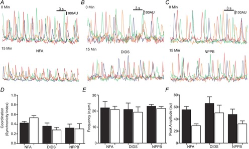
Figure 4. No further effects on Ca2+ transients with pharmacological inhibitors were observed in Ano1 KO mice.

A–C, representative traces of Ca2+ transients upon pharmacological agent treatment in Ano1 KO tissue: 15 min treatment with NFA (1 μm, A), DIDS (10 μm, B) and NPPB (10 μm, C). D, measurements in Ano1 KO tissues upon treatment with NFA, DIDS and NPPB showed no significant difference in coordination of the events as indicated by the synchronicity index (n = 3, P = n.s., paired t test). E, no significant difference in frequency was observed in Ano1 KO tissue upon treatment with NFA, DIDS and NPPB (n = 3, P = n.s., paired t test). F, no significant difference in peak amplitude was observed in Ano1 KO tissue upon treatment with NFA, DIDS and NPPB (n = 3, P = n.s., paired t test).
