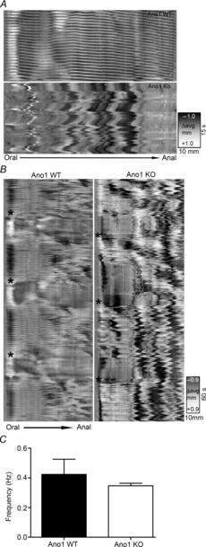
Figure 9. Altered spatiotemporal maps in Ano1 KO mice small intestines.

Representative images of patterns of motility from Ano1 WT and Ano1 KO mouse small intestinal preparation. A, the spatiotemporal maps (top) from an Ano1 WT mouse exhibiting regular and rhythmic contractions (referred to as ‘ripples’) whereas in Ano1 KO (bottom) showed absence of ripple activity signifying loss of rhythmic synchronized contractile activity. B, representative spatiotemporal maps on a lower scale from Ano1 WT mice (left) again showing ripples and also showing a phasic modulation of contractility across the entire preparation, probably corresponding to synchronized contractile activity (black asterisks). The right panel again shows uncoordinated ripples and shows that phasic modulation of contractile activity still appeared to be present (black asterisks) in Ano1 KO preparations. C, spatiotemporal map analysis revealed no significant difference in frequency of contractions in Ano1 WT and Ano1 KO mice (mean ± SEM, n = 3, P = n.s., unpaired t test).
