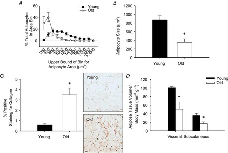
Figure 1. Epididymal white adipose tissue (eWAT) adipocyte area, tissue fibrosis and adipose tissue volumes in young and old mice.

Adipocyte area was determined by measuring cross-sectional area of adipocytes (521 ± 71 adipocytes per animal; range: 211–800) on haematoxylin and eosin stained histological sections of eWAT from young (n = 8) and old (n = 7) mice and are presented as a histogram of adipocyte area bins (A) and as mean area (B). C, fibrosis was assessed by picrosirius red staining for collagen in histological sections of eWAT from young (n = 6) and old (n = 8) mice. Representative images shown to the right of the summary data. D, visceral and subcutaneous adipose tissue volume was assessed in young (8 months, n = 5) and old (25 months, n = 4) mice by CT and expressed relative to total body mass. *Denotes group difference from young. Data are means ± s.e.m., P ≤ 0.05.
