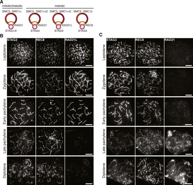
Figure 1. Chromosomal localization of meiotic cohesin components.
A Composition of different cohesin complexes during mitosis and meiosis.
B, C Chromosomal localization of STAG3 during prophase I. Nuclear spreads of wild-type spermatocytes were immunostained for STAG3, REC8, and RAD21L (B) or for STAG3, REC8, and RAD21 (C). Note that the late pachytene spermatocyte in (C) is at a later stage than the one in (B). Bars, 10 μm.

