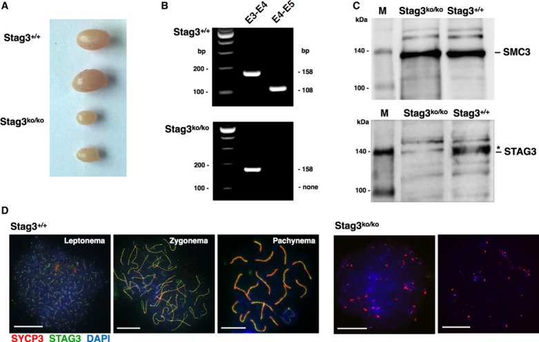
Figure 1. Characterization of spermatogenesis in Stag3ko/ko mice.

- Testis samples from wt (Stag3+/+) and Stag3ko/ko mice, 40 days of age.
- RT-PCR analysis of testis mRNA from wt (Stag3+/+) and Stag3ko/ko mice. The primer pairs are shown indicating the respective exons (E3, E4, E5); the expected size (bp) of the PCR products is provided.
- Immunoblot of testis nuclear extracts of the indicated mice, probed with anti-STAG3 or anti-SMC3 antibody as indicated. The anti-STAG3 antibody recognizes a specific band corresponding to the predicted molecular weight (141 kDa) of STAG3, which was present in wt but absent in Stag3ko/ko extracts. An unspecific band is marked by an asterisk. A gel was loaded in parallel using the same extracts, and the corresponding membrane was probed with an antibody directed against SMC3, which has the same predicted molecular weight (141 kDa). The pictures are representative of three independent experiments. M = biotinylated protein marker.
- Immunofluorescence staining of spermatocyte chromosome spreads of wt and Stag3ko/ko mice, probed with anti-SYCP3 antibody for AEs and SCs and anti-STAG3; nucleic acids were stained with DAPI. The stages of wt prophase I spermatocytes are indicated, and two examples of Stag3ko/ko chromosome spreads are provided. Size bars indicate 10 μm.
Source data are available online for this figure.
