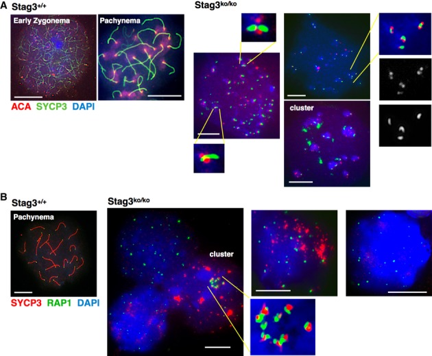
Figure 4. Centromeres and telomeres in Stag3ko/ko spermatocytes.

- Immunofluorescence staining of spermatocyte chromosome spreads of wt and Stag3ko/ko mice, probed with anti-SYCP3 and anti-centromere antibody (ACA); nucleic acids were stained with DAPI. Three examples of Stag3ko/ko spreads are shown to indicate centromere cluster formation and to highlight partial co-localization of SYCP3 with centromeres and the structures of these regions. Three areas are provided as magnified excerpts. Size bars indicate 10 μm.
- Immunofluorescence staining of spermatocyte chromosome spreads of wt and Stag3ko/ko mice, probed with anti-SYCP3 and anti-RAP1 to stain telomeres; nucleic acids were stained with DAPI. Three examples of Stag3ko/ko spreads are shown to represent different stages and to show an example of telomere cluster formation, which is shown in a magnified excerpt as well. Size bars indicate 10 μm.
