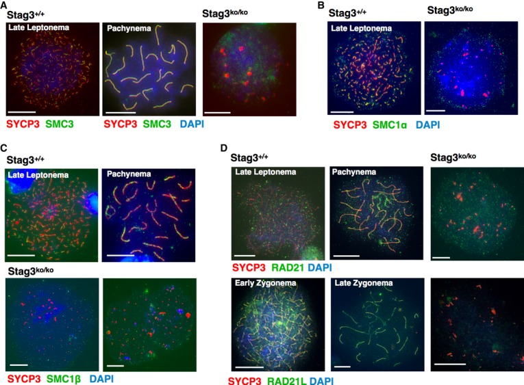
Figure 5. Cohesin proteins in Stag3ko/ko spermatocytes.

A–D Immunofluorescence staining of spermatocyte chromosome spreads of wt and Stag3ko/ko mice, probed with (A) anti-SYCP3 and anti-SMC3; (B) anti-SYCP3 and anti-SMC1α; (C) anti-SYCP3 and anti-SMC1β; (D) anti-SYCP3 and anti-RAD21 or anti-RAD21L. Nucleic acids were stained with DAPI. Size bars indicate 10 μm.
