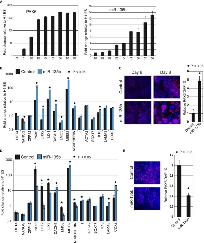
Figure 4. MiR-135b promotes neuroectodermal fate in human pluripotent cells.
- Time course of miR-135b and PAX6 expression during neural differentiation of H1 ESC. MiR-135b levels were quantified by Taqman assays.
- Quantitative RT-PCR data to measure gene expression changes after miR-135b transfection. Fold changes were compared to untransfected H1 ESC.
- Immunostaining for PAX6 in miR-135b-transfected H1 ES cells at day 6 and day 8 after induction of differentiation.
- Quantitative RT-PCR data to assay gene expression after miR-135b knockdown in H1 ESC. Fold changes were compared to untransfected H1 ESC.
- Immunostaining for PAX6 after miR-135b knockdown in differentiated H1 ESC at day 8.
Data information: All error bars shown indicate s.e.m. (n = 3 independent experiments). Images were quantified as described in Materials and Methods. P-values indicated were calculated using the Student's one-tailed t-test.

