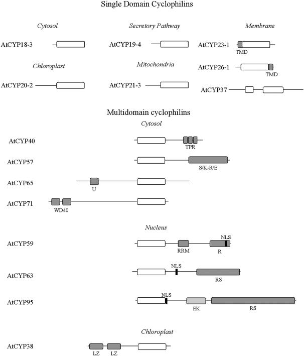
Figure 1.
Primary domain structure of representative isoforms of the SD and MD Arabidopsis cyclophilin proteins. TMD, transmembrane domain; TPR, tetratricopeptide repeat; S/K-R/E, Ser/Lys-Arg/Glu-rich region; U, U-box domain; WD40, WD40 repeat; RRM, RNA recognition motif; R, Arg- rich region; NLS, nuclear localization signal; EK, Glu-Lys-rich region; RS, Arg-Ser rich region; LZ, Leu zipper.

