
Figure 4. Treg-mediated suppression requires Ikaros activity.
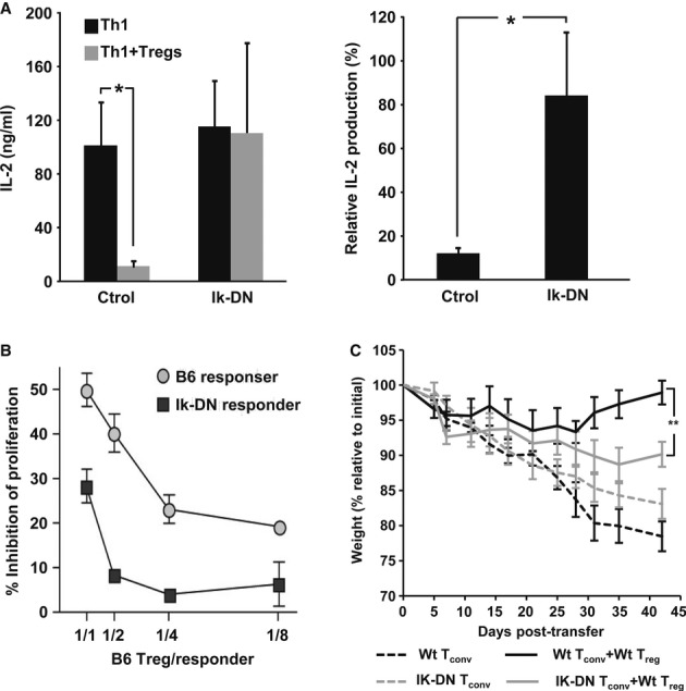
A Th1 cells were transfected with a plasmid expressing IK-DN or a control vector and activated in the presence of Tregs (1:1). Percent suppression of IL-2 expression was calculated (mean + s.e.m. from four experiments, t-test, *P < 0.05).
B B6 or B6-IkDN/+ CD4+CD25− T cells (Thy1.2+) were labeled with CFSE and activated with APC and soluble anti-CD3 in the presence of increasing amounts of CD4+CD25+ Treg from B6-Thy1.1+ mice. After 3 days, suppression of responder cell proliferation was determined by FACS, assessing the degree of inhibition of CFSE dilution (mean ± s.e.m.) from two experiments.
C CD25−CD4+ Tconv cells from WT or IK-DN B6 mice were transferred into B6-Rag1−/− mice. Twenty days later, mice received PBS or CD25+ CD4+ Treg purified from WT donors. Recipients were weighed (mean ± s.e.m.) and observed for symptoms of diarrhea approximately every 2 days (5 mice per group. one-way ANOVA **P < 0.01).
Source data are available online for this figure.