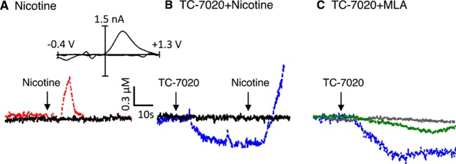
Figure 1.

Representative voltammetric recordings of real-time dopamine signaling in nucleus accumbens of anaesthetized rats before and after administration (arrows) of (A) nicotine (0.3 mg/kg iv), (B) TC-7020 (1 mg/kg iv) followed by nicotine, and (C) TC-7020 alone (blue trace) or TC-7020 after pretreatment with MLA (10 mg/kg ip) (green trace) or MLA alone (black trace). Black traces in A and B are controls after saline injection. Inset to panel A is representative background-subtracted voltammogram obtained at peak of response, showing characteristic oxidation and reduction peak potentials (approximately +0.6 V and approximately −0.2 V, respectively) that identify dopamine.
