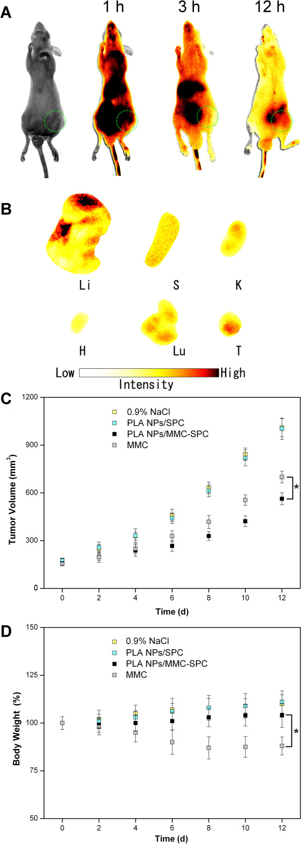
Figure 8.
In vivo fluorescence imaging and in vivo anticancer effect of the hybrid PLA NPs/MMC-SPC. (A)In vivo fluorescence imaging of H22 tumor-bearing mice after intravenous injection of the DiR-PLA NPs/MMC-SPC at 1, 3, and 12 h post-injection. (B)Ex vivo fluorescence imaging of H22 tumor-bearing mice after intravenous injection of the DiR-PLA NPs/MMC-SPC at 12 h post-injection (Li, liver; S, spleen; K, kidney; H, heart; Lu, lung; T, tumor). (C) The change of tumor volumes of H22 tumor-bearing mice treated with PBS, PLA NPs/SPC, PLA NPs/MMC-SPC, and MMC. (D) The change of body weights of H22 tumor-bearing mice treated with 0.9% NaCl, PLA NPs/SPC, PLA NPs/MMC-SPC, and MMC.

