Figure 3.

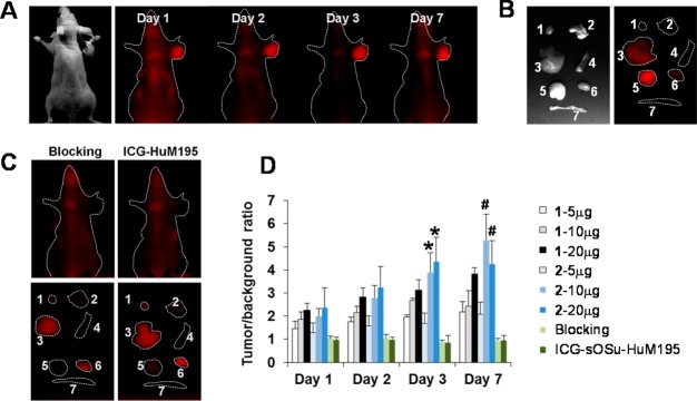
(A) Representative in vivo NIR fluorescence imaging of athymic mice bearing HER1-positive s.c. LS-174T xenografts at days 1, 2, 3, and 7 after i.v. injection of SE-HPLC-purified ICG-sOSu-panitumumab (2, 20 μg). (B) Representative ex vivo NIR fluorescence image (right) of the dissected organs at day 3. Labels: 1, heart; 2, lung; 3, liver; 4, spleen; 5, tumor; 6, kidney; 7, intestine. Note: The white light image in (B) indicates the order of the organs shown in (B) and (C). (C) Representative in vivo and ex vivo fluorescence images of HER1-positive s.c. LS-174T tumor-bearing mice with excess antibody blocking or with ICG-sOSu-HuM195 (negative control). (D) Comparison of tumor-to-background ratio with injection of various doses of SE-HPLC-purified ICG-sOSu-panitumumab (1, 2) in athymic mice bearing HER1-positive s.c. LS-174T xenografts (n = 5). *, significantly different from 2 - 5 μg group (day 3) (P < 0.001); #, significantly different from 2 - 5 μg group (day 7) (P < 0.01).
