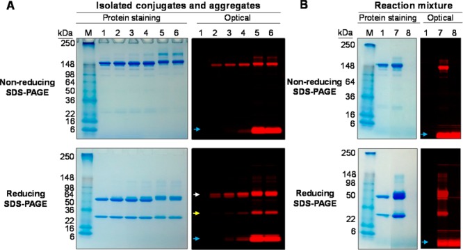
Figure 4.

SDS-PAGE of (A) SE-HPLC-isolated ICG-sOSu-panitumumab conjugates and aggregates, and (B) reaction mixture, under nonreducing and reducing conditions with β-mercaptoethanol. Panitumumab served as a reference. Colloidal blue protein staining and optical imaging were performed: M, marker; 1, intact panitumumab antibody; 2, 1; 3, 2; 4, 3; 5, ICG-sOSu aggregates (from 20× reaction, peak with RT = 12.5 min); 6, ICG-sOSu aggregates (from 20× reaction, peak with RT = 14.5 min); 7, ICG-sOSu-panitumumab reaction mixture (10×, without purification); 8, ICG-sOSu alone incubated in conjugation buffer for 1 h at 37 °C as a control. White arrow: heavy chain; yellow arrow: light chain; blue arrow: free dye.
