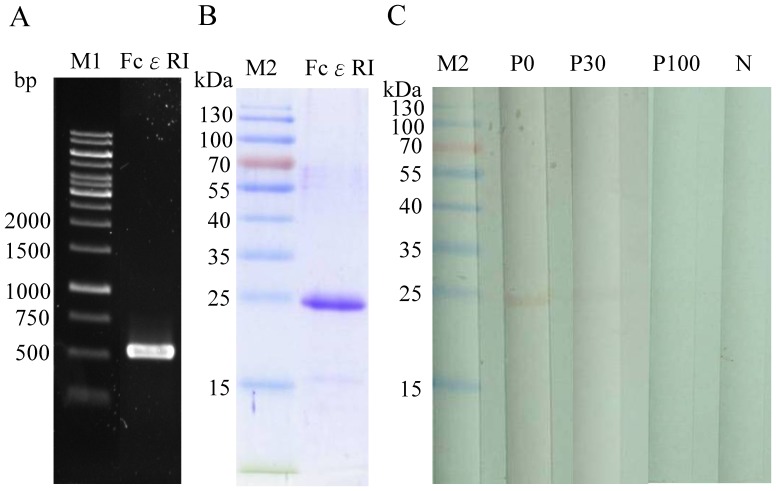
Figure 1. Analysis of cDNA and E. coli-expressed protein of FcεRIα.
(A) Agarose gel electrophoresis of amplified cDNA coding for FcεRIα (534 bp) obtained by RT-PCR from total RNA of human basophils. (B) Coomassie blue-stained SDS-PAGE and (C) immuno-blotting and immunoblot inhibition of purified recombinant FcεRIα protein with pooled sera from CU-ASST(+) and CU-ASST(-) subjects. Lane P0 was pooled CU-ASST(+) patient sera immunoblotted with purified recombinant FcεRIα protein, lanes P30 and P100 showed pooled CU-ASST(+) sera pre-incubation with 30 or 100 µg/ml of rFcεRIα proteins. Lane N was sera from pooled CU-ASST(-) patients. Numbers on the left indicate sizes of DNA (M1) and protein markers (M2).

