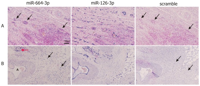
Figure 3. MiR-664-3p in situ hybridization (ISH).
Panels A and B show examples of miR-664-3p ISH in infiltrating lymphocytes (A) and fibroblasts (B). Consecutive sections were stained with LNA probes against miR-664-3p, miR-126-3p and a scramble sequence. MiR-664-3p ISH signal is seen in infiltrating lymphocytes (A, arrows; B, red arrow) and in fibroblasts (B, black arrows), whereas no ISH signal is obtained with scramble probe. A strong ISH signal is seen in endothelial cells with the positive control probe miR-126-3p. The “A” in panel B indicates an artery.

