Figure 1.

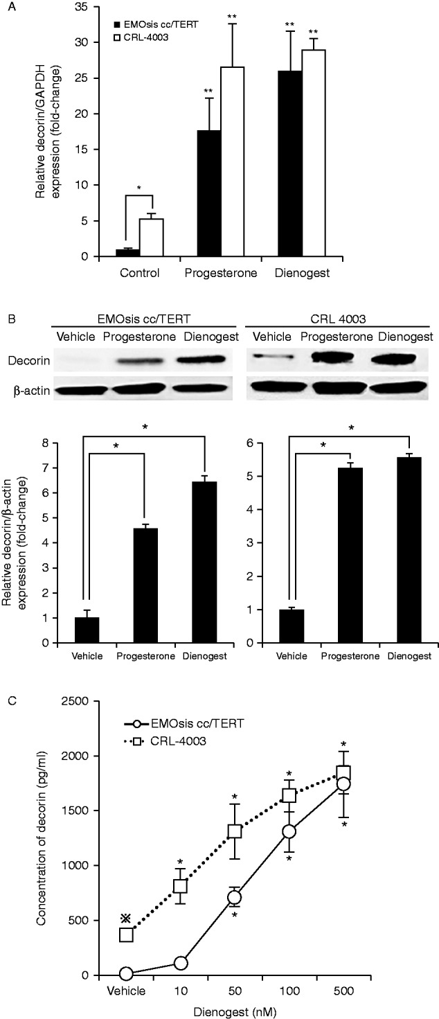
(A) Progesterone treatment promoted DCN mRNA expression in EMOsis cc/TERT cells and CRL-4003 cells. The mRNA expression of decorin was determined by real-time PCR from the total RNA obtained from CRL-4003 cells and EMOsis cc/TERT cells cultured with or without decorin or dienogest for 12 h. The mRNA levels of decorin were normalized to those of GAPDH mRNA. The data were processed by the comparative Ct method and expressed as a fold-increase relative to the basal transcription level in the control. Bars indicate the s.e.m. Significant differences are indicated by asterisks. *P<0.05, **P<0.01. (B) Progesterone treatment promoted the expression of decorin in EMOsis cc/TERT cells and CRL-4003 cells. CRL-4003 cells and EMOsis cc/TERT cells were harvested and used to prepare cell lysates after treatment with or without dienogest or progesterone for 24 h. The lysates were subjected to SDS–PAGE and blotted with anti-decorin (upper panel) or anti-β-actin (lower panel) antibodies. The lower panel shows the densitometric quantification of the western blot analysis normalized to β-actin expression and expressed as a fold-increase relative to the basal transcription level in the control. The mean ±s.d. of three determinations is shown. *P<0.05. (C) Progesterone treatment promoted the secretion of decorin in EMOsis cc/TERT cells and CRL-4003 cells. CRL-4003 cells and EMOsis cc/TERT cells were treated with dienogest at various concentrations for 48 h, and the concentrations of decorin produced by the EMOsis cc/TERT and CRL-4003 cells were measured using the Decorin (DCN) Human ELISA kit. The data are expressed as the mean±s.d. (N=5). * indicates a significant (P<0.05) difference compared with the untreated control group, while ⋇ indicates a significant (P<0.05) difference compared with untreated control EMOsis cc/TERT cells.

 This work is licensed under a