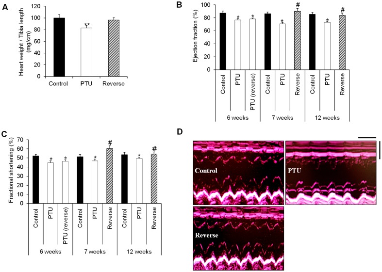
Figure 6. The LT4 treatment reversed cardiac dilation and improved systolic function.
A: Heart weight to tibia length ratio (mg/cm) in the different treatment groups. Statistical analysis was performed with Kruskal-Wallis One-way ANOVA on rank tests followed by post hoc Dunn's multiple comparison tests. **p<0.01 vs. control and reverse. B and C: Echocardiographic measurements of the ejection fraction (%) and fractional shortening (%) in the control, PTU-treated and LT4-treated rats at different time points. Statistical analysis was performed with two-way ANOVA tests followed by post hoc Tukey's multiple comparison tests. *p<0.05 vs. control as well as 7 and 12 weeks reverse; # p<0.05 vs. PTU. D: M-mode echocardiographic tracings from the control, PTU and reverse groups. Echocardiography scale bars: 0.25 s and 0.5 mm. Data are represented as the mean ± SEM. n = 10 animals for each group.

