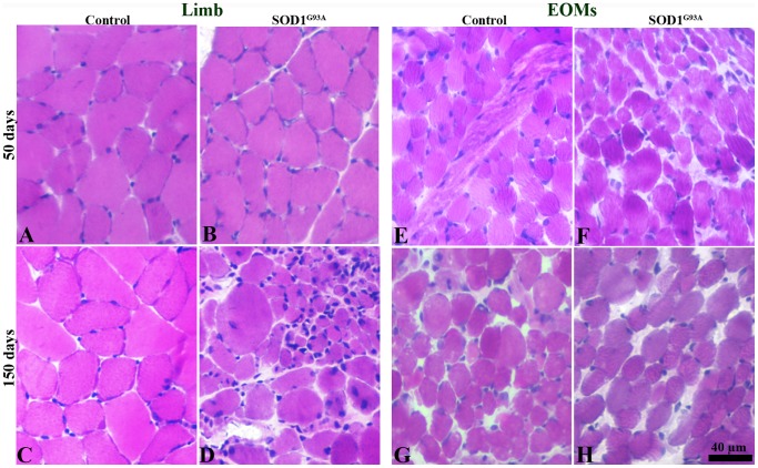
Figure 1. H&E staining of limb muscles (A–D) and EOMs (E–H) from control and SOD1G93A mice at 50 days and 150 days.
Normal morphological features of muscle fibres in limb muscles (B) and EOMs (F) at 50 days and in EOMs (H) at 150 days. In limb muscles of terminal stage SOD1G93A mice (D), muscle fibres presenting pathological alterations including atrophy, hypertrophy, round fibres, and fibres with central nuclei.

