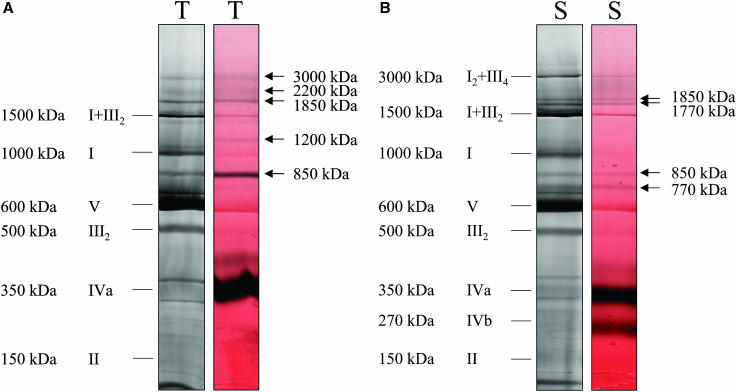
Figure 1.
Identification of complex IV-containing supercomplexes in potato tuber (T) and stem (S) mitochondria. Protein complexes were solubilized by 5 g digitonin per g protein, separated by 1D BN-PAGE and either visualized by Coomassie staining (left gel strips) or by in-gel activity staining for cytochrome c oxidase (right gel strips). Activity stains are given in false-color mode to increase color contrast (red, Coomassie; black, enzyme activity). Molecular masses and identities of known protein complexes are indicated on the left side of the gels in Roman numerals (I, NADH dehydrogenase; II, succinate dehydrogenase; III, cytochrome c reductase; IVa and IVb, large and small form of cytochrome c oxidase; V, ATP synthase; I + III2 and I2 + III4, supercomplexes of complexes I and III). Additional supercomplexes exhibiting cytochrome c oxidase activity are indicated by arrows.

