Figure 2).

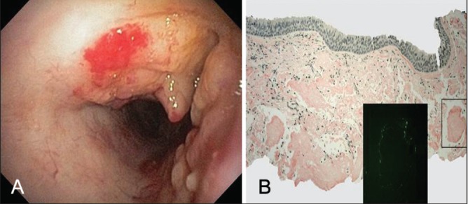
Bronchoscopic view of mid trachea showing nodular thickening with cobblestone appearance and luminal narrowing, relatively sparing the membranous portion (A). Tracheal biopsy showing submucosal amyloid deposits staining positive with Congo red (original magnification ×40) and apple-green birefringence under polarized light (original magnification ×200) (B)
