
Figure 1.
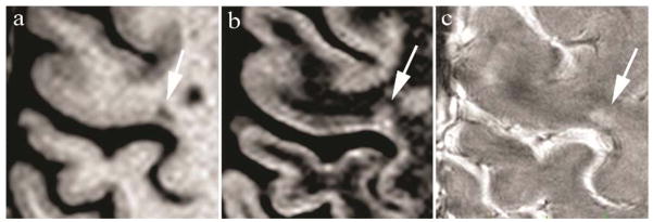
Prospective scoring on 3T MEMPR (a) and DIR (b) disclosed a juxtacortical lesion (arrow). Prospectively, 7T FLASH-T2* (c) confirmed the lesion as a type I (arrow). Retrospective analysis re-categorized the juxtacortical lesion (a, b) as a type I CL. Time between scans is 6 days.