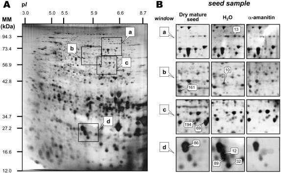
Figure 5.
Influence of α-amanitin on the proteome of tt2-1 Arabidopsis mutant seeds 24 h after sowing. An equal amount (200 μg) of total protein extracts was loaded in each gel. A, Silver-stained 2D gel of total proteins from tt2-1 seeds incubated for 24 h in water. The indicated portions of the gel, a, b, c, and d, are reproduced in B. B, enlarged windows (a–d) of 2D gels as shown in A for tt2-1 dry mature seeds (left), tt2-1 dry mature seeds incubated in water for 24 h (middle) and tt2-1 dry mature seeds incubated in 500 μm α-amanitin for 24 h (right). The nine labeled protein spots (protein nos. 13, 161, 10, 194, 69, 86, 89, 12, and 32) were identified by comparison with Arabidopsis seed protein reference maps (Gallardo et al., 2001, 2002a, http://seed.proteome.free.fr; see Table II). Protein spot quantitation was carried out as described in “Materials and Methods,” from at least three gels for each seed sample.

