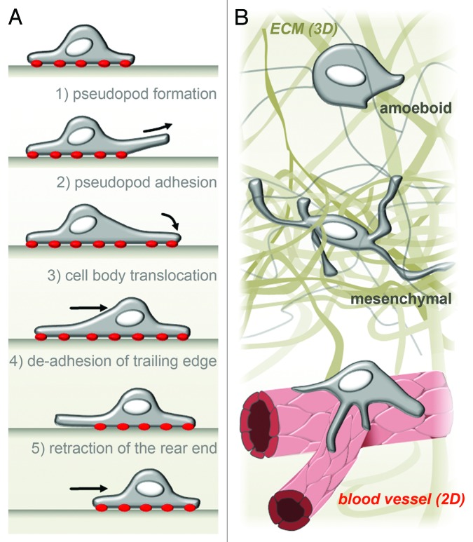
Figure 1. Macrophage migration in 2D and 3D. (A) In vitro, on 2D surfaces, human macrophages adopt a rounded, flat cell shape and follow a classical five-step model of cell migration. Adhesion to the substrate is marked by red dots. (B) In vivo, macrophages are confronted with both 2D (B, bottom) and 3D (B, middle and top) situations. For example, during vascularization, macrophages can be found on vascular junctions attached to a 2D endothelial wall. Depending on the extracellular environment, macrophages migrating through interstitial space can adopt a rounded cell shape and use the amoeboid, non-proteolytic migration mode (B top) or a prolonged protrusion-rich morphology and use mesenchymal, proteolysis-dependent migration (B middle).

An official website of the United States government
Here's how you know
Official websites use .gov
A
.gov website belongs to an official
government organization in the United States.
Secure .gov websites use HTTPS
A lock (
) or https:// means you've safely
connected to the .gov website. Share sensitive
information only on official, secure websites.
