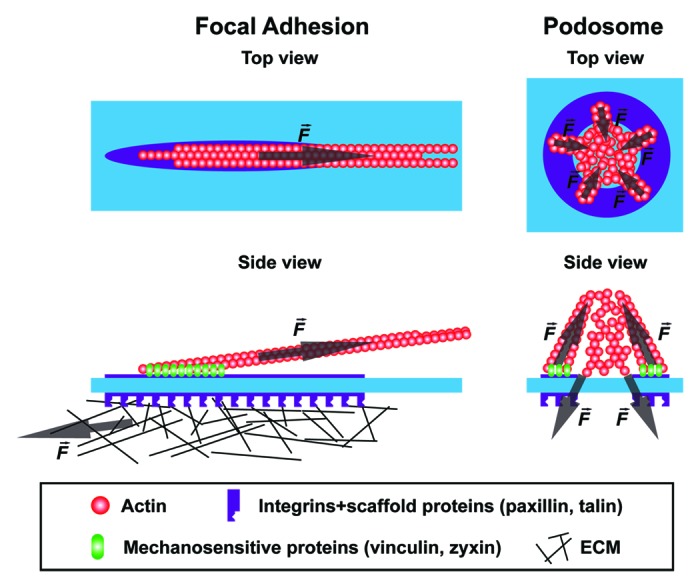
Figure 1. Force distribution within podosomes and focal adhesions. Schematic representation of the top and side view of a focal adhesion and a podosome. The forces generated within these adhesions are indicated with an arrow. The growth of focal adhesions and podosomes is both tension-mediated but since opposite forces are generated within a podosome, force derived from integrin-mediated ECM ligation is not necessary to facilitate the growth of podosomes.
