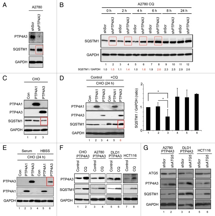
Figure 4. PTP4A3 promotes SQSTM1 degradation and serves as a substrate in autophagy. (A) A2780 cells were infected with stable shRNA knockdown constructs against nonspecific targets (shScr) or PTP4A3 (shPTP4A3). Exponentially growing A2780-shScr and A2780-shPTP4A3 cells were lysed and analyzed for stable-state SQSTM1 and PTP4A3 expression levels by western blotting. (B) A2780-shScr and A2780-shPTP4A3 cells were treated with CQ for indicated times before lysis and western blotting analysis of SQSTM1 expression levels. The band intensity ratio of SQSTM1/GAPDH was quantified as described in Materials and Methods, and presented as a histogram in the right panel (mean ± S.D.). (C) CHO-Con, CHO-PTP4A1, and CHO-PTP4A3 stable cells were analyzed for stable-state SQSTM1 expression levels by western blotting. (D) CHO-Con, CHO-PTP4A1, and CHO-PTP4A3 cells were cultured in full medium in the absence (control) or presence (CQ) of CQ for 24 h before lysis and western blot analysis of the indicated proteins. The ratio of SQSTM1/GAPDH was quantified as described in Materials and Methods, and presented as a histogram in the right panel (mean ± S.D.). (E) CHO-Con, CHO-PTP4A1, and CHO-PTP4A3 cells were cultured in full media (control) or in HBSS for 24 h prior to lysis and western blotting analysis of the indicated proteins. (F) CHO-PTP4A3, A2780-PTP4A3, and HCT116 cell lines were cultured in full media in the absence (control) or presence (CQ) of CQ for 24 h prior to lysis and western blotting analysis of the indicated proteins. (G) ATG5 was knocked down using shRNA in A2780-PTP4A3 and HCT116 cells. Exponentially growing cells (in full media) were lysed for western blotting analysis with the indicated antibodies. GAPDH was used as loading control.

An official website of the United States government
Here's how you know
Official websites use .gov
A
.gov website belongs to an official
government organization in the United States.
Secure .gov websites use HTTPS
A lock (
) or https:// means you've safely
connected to the .gov website. Share sensitive
information only on official, secure websites.
