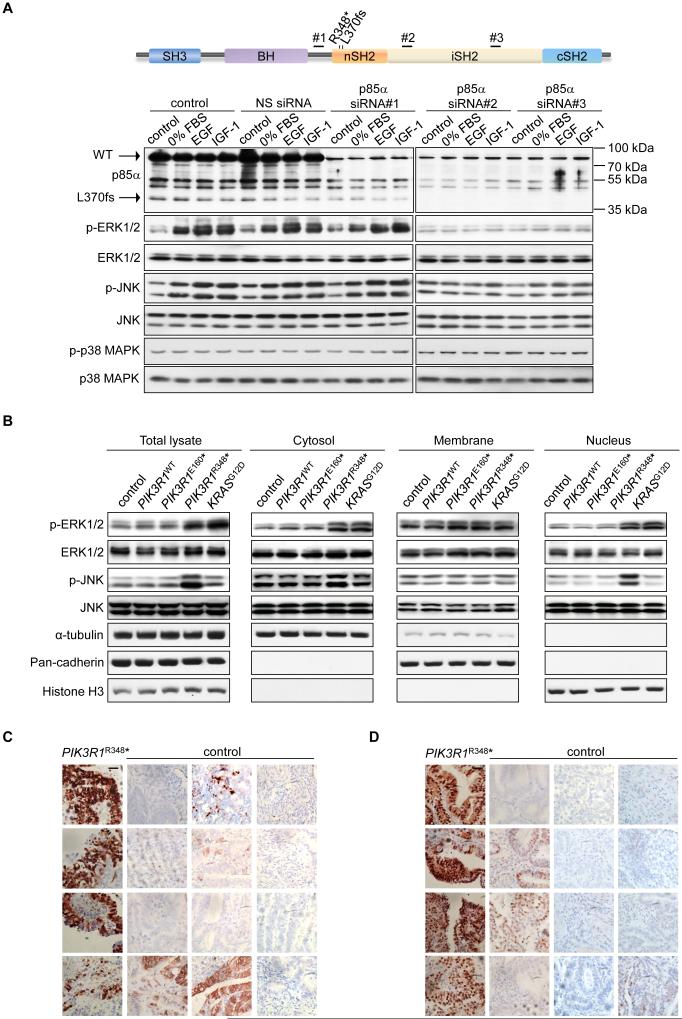
Figure 2. PIK3R1R348*- and PIK3R1L370fs-mutant cell lines and endometrial tumors show high levels of p-ERK and p-JNK.
(A) Top, schematic showing the locations of p85α siRNA-targeted regions; Bottom, ovarian endometrioid cancer cells OVK18 transfected with p85α siRNAs or non-specific (NS) control for 72 hr were cultured in the absence of FBS (0% FBS) for additional 24 hr. The cells were then simulated with epidermal growth factor (EGF; 10 ng/ml) or insulin-like growth factor 1 (IGF-1; 10 ng/ml) for another 1 hr before being harvested for Western blotting. Lysates were loaded into two gels due to sample well limits but the membranes were probed with same antibodies for equal duration and the proteins were detected under same exposure time. (B) Western blotting of total lysates or subcellular fractions from SKUT2 endometrial cancer cells transfected with PIK3R1WT, PIK3R1E160*, PIK3R1R348* or KRASG12D. α-tubulin (cytosolic), pan-cadherin (membrane) and histone H3 (nuclear) served as markers for purity of fractions. (C, D) Immunohistochemical staining for p-ERK (C) and p-JNK (D) in 4 endometrial carcinomas carrying PIK3R1R348* and 12 endometrial carcinomas without PIK3R1R348* (control). Nuclei were counterstained in hematoxylin. Bar, 50 μm. See also Figure S2 and Table S4.

