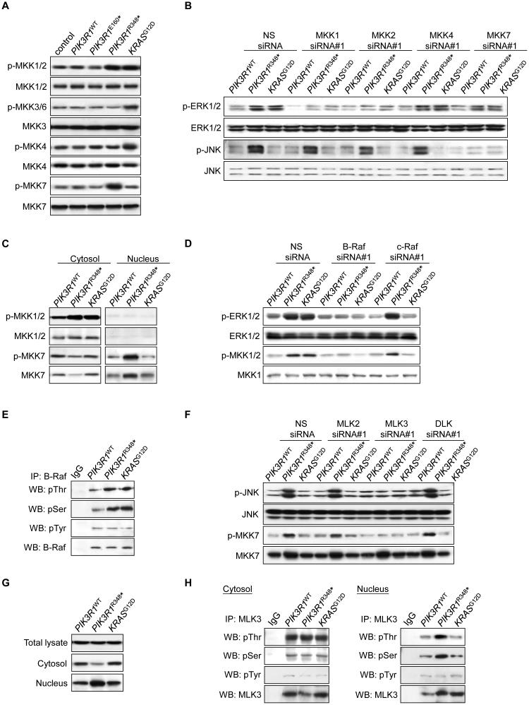
Figure 3. PIK3R1R348* selectively activates B-Raf-MKK1/2-ERK and MLK3-MKK7-JNK pathways.
(A) Total lysates from transfected SKUT2 were analyzed by Western blotting. (B) Cells co-transfected with PIK3R1WT, PIK3R1R348* or KRASG12D and 20 nM siRNAs targeting the MKKs or non-specific (NS) control for 72 hr were harvested for subcellcular fractionation. Western blots shown were from nuclear lysates. (C) Transfected SKUT2 were harvested for subcellcular fractionation and Western blotting. (D) Cells were co-transfected with PIK3R1WT, PIK3R1R348* or KRASG12D and 10 nM siRNAs targeting B-Raf, c-Raf or NS control for 72 hr. Data shown are Western blots of cytosolic lysates. (E) Total lysates from transfected SKUT2 were harvested for immunoprecipitation (IP) with anti-B-Raf antibody and Western blotting (WB). IP with anti-IgG was used as control. (F) Cells co-transfected with PIK3R1WT, PIK3R1R348* or KRASG12D and 20 nM siRNAs targeting MLK2, MLK3, DLK (40 nM) or NS control for 72 hr were harvested for subcellular fractionation. Western blots shown were from nuclear lysates. (G) Western blots for MLK3 in total lysates, cytosolic and nuclear fractions from transfected SKUT2. (H) Western blots of IP with anti-MLK3 antibody using cytosolic and nuclear extracts from transfected SKUT2. See also Figure S3

