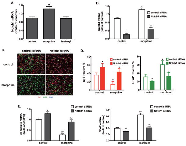
Figure 2. Morphine promotes astrocyte-preferential differentiation via Notch1.
(A) The expression of Notch1 was determined by real-time PCR after 4 d of differentiation, in the presence of 1 μM morphine or 10 nM fentanyl. The results were normalized against those of GAPDH, and further normalized against the result obtained from the control group. *, p<0.05 compared to control.
(B) The expression of Notch1 was determined by real-time PCR after transfection with control and Notch1 siRNA. The results were normalized against those of GAPDH. **, p<0.01 compared to control siRNA transfected group with the same treatment; #, p<0.05 compared to the control siRNA transfected group without morphine treatment.
(C) Adult hippocampus-derived neural progenitor cells were transfected with control siRNA or Notch1 siRNA, and cultured in complete differentiation medium with 1 μM morphine for 4 d. Cells were stained with markers for neurons (Tuj1), astrocytes (GFAP) and with DAPI. Scale bar, 25 μm. Images are representative of at least three independent experiments with similar results.
(D) Quantification of cells stained with each marker, calculated as the percentage of the total number of cells stained with DAPI. Red: Tuj1; Green: GFAP. *, p<0.05 compared to the control siRNA group with the same treatment; #, p<0.05 compared to the control group transfected with control siRNA.
(E) The expression of βIII-tubulin and GFAP were determined by real-time PCR after 4 d of differentiation with indicated treatments. The results were normalized against those of GAPDH. *, p<0.05, **, p<0.01, compared to control-siRNA-transfected group with the same treatment. ##, p<0.01 compared to the control group transfected with control siRNA. All data represent mean ± SEM of four independent experiments.

