
Figure 4. Western blots.
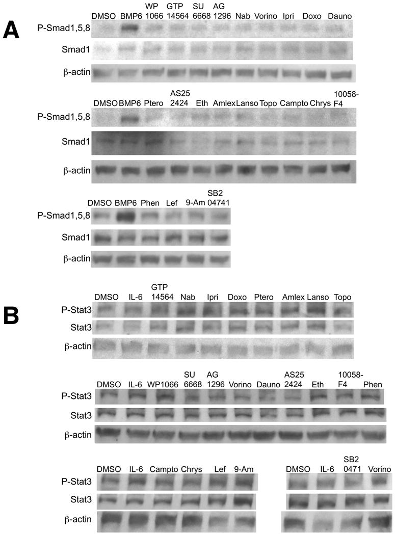
Following 16 hours of serum starvation in α-MEM/1%FBS, HepG2 cells were treated for 1 hour in α-MEM/1%FBS with the chemicals at the same concentrations as given in Figure 2. Protein was extracted from the cells, separated by SDS-PAGE, and blotted for incubation with antibodies against anti-P-Smad1,5,8 (A) or P-Stat3 (B). Following immunoblotting, the membranes were stripped and re-probed with either antibody against anti-Smad1,5,8 (A) or Stat3 (B). The blots were then stripped again and probed for β-actin as a loading control. Each chemical was evaluated in two or three biological replicates.