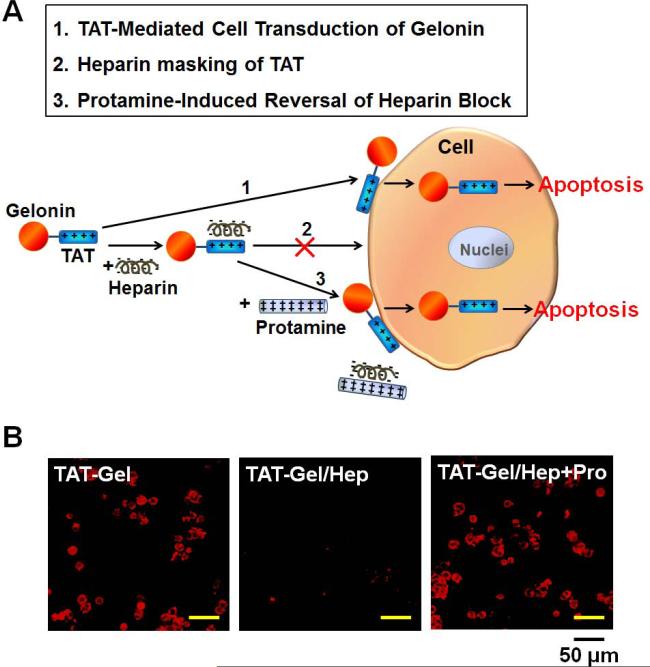
Figure 6.
Heparin/protamine modulation of TAT-Gel cell transduction. (A) Schematic illustration of regulating TAT-Gel cell transduction via reversible masking and demasking of TAT on the TAT-Gel by anionic heparin and cationic protamine, respectively. (B) Confocal microscopic images of LS174T cells treated with TATGel, TAT-Gel/Hep or “TAT-Gel/Hep+Pro”. LS174T cells were plated onto 8-well chambered coverglass (Thermo Scientific, Rockford, IL) at a density of 105 cells/well and, when cells were attached to the bottom of the chambers, TRITC-labeled TAT-Gel (5 μM) was added to the wells either 1) alone, 2) with heparin (TAT-Gel/Hep) or 3) with heparin and protamine (“TAT-Gel/Hep+Pro”). After 3 h incubation, cells were washed and the live cell images were acquired by a Nikon A1R-A1 confocal laser microscope with a 20×objective. The TAT-Gel/Hep complex was prepared by mixing TAT-Gel with 3-fold molar excess of heparin and incubation at 4°C for 30 min. For “TAT-Gel/Hep+Pro” treatment, cells were treated with TAT-Gel/Hep (with 3-fold molar excess of heparin to TAT-Gel), followed by immediate addition of protamine (3-fold molar excess against heparin). (TAT-Gel: recombinant TAT-gelonin fusion protein)

