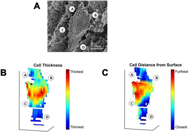
Figure 9.
3D reconstructions of a wild type MG63 cell grown on SLA (A), a color gradient indicating its cell thickness (B), the same color gradient indicating the distance between the cell and substrate surface (C). The reconstructions in (B) and (C) are viewed from a top-down perspective, as for the cell shown in (A). The locations indicated by A, B, C and D for the imaged cell in (A) are also indicated in the 3D reconstructions in (B) and (C).

