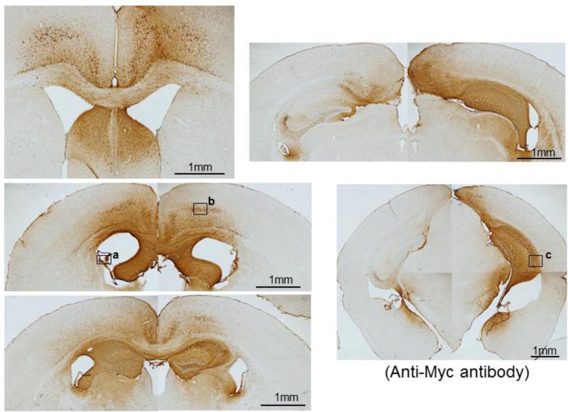
Fig 3. IgVL5D3 is predominantly expressed in the right brain hemisphere.
rAAV9-IgVL5D3 was injected into the right lateral ventricle of TgAPPswe/PS1dE9 mice and, 8 (prophylaxis) and 5 (therapy) months after the injection, IgVL5D3 expression in the brain was visualized by immunohistochemistry using anti-c-Myc antibody. Brain sections from a representative mouse are shown. Scale bars, 1 mm. High magnification pictures of the areas indicated by the squares (a through c) are found in Supplemental Figure 1.

