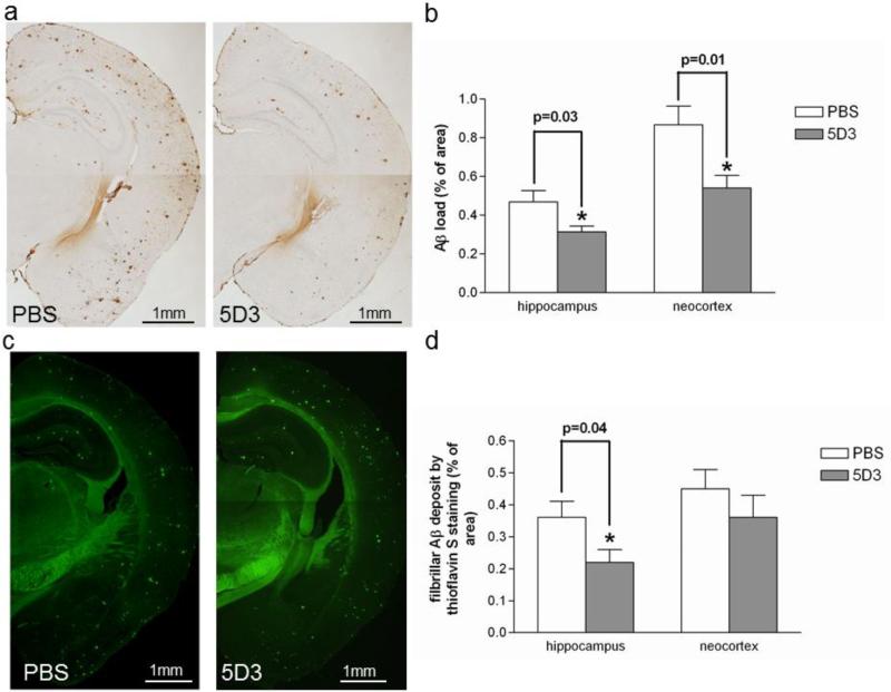
Fig 5. Prophylactic rAAV9-IgVL5D3 injection decreases Aβ load in the right hippocampus.
Eight months after rAAV9 or PBS injection, TgAPPswe/PS1dE9 mice were terminated and Aβ deposits in the brain were visualized and quantified by morphometric analysis. Aβ deposits are visualized by 6E10 antibody (a, b) and thioglavine S (c, d). The percentages of immunoreactive or fluorescent areas for 6E10 (b) and thioflavin S (d), respectively, are shown as bar graphs. The values shown are the mean ± SEM. PBS: PBS-injected mouse, 5D3: rAAV9-IgVL5D3-injected mouse. Scale bars, 1 mm.

