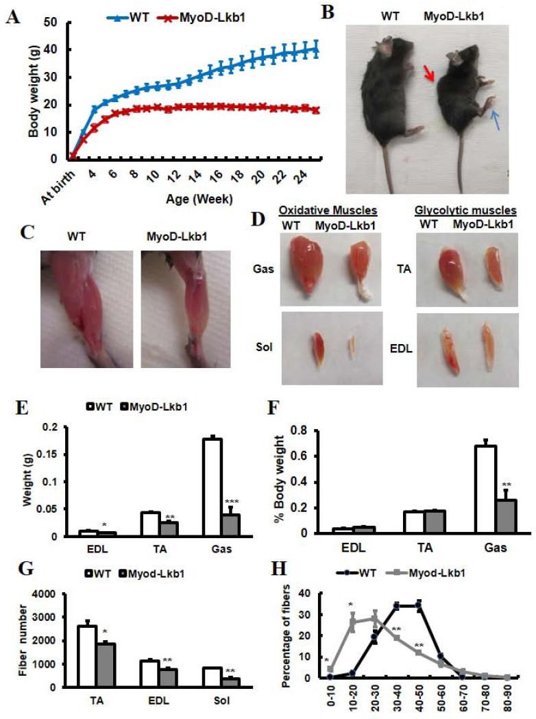
Figure 1.
MyoDCre-mediated deletion of Lkb1 impairs muscle development and growth. (A) Growth curve of the WT (Lkb1flox/flox) and MyoD-Lkb1 (MyoDCre/ Lkb1flox/flox) mice. P < 0.01 for all timepoints measured, n=8 for WT and n=5 for MyoD-Lkb1 mice. (B) Gross morphology of WT and MyoD-Lkb1 mice at 10-wk-old. Kyphosis (red arrow) and abnormal hindlimb postures (blue arrow) were indicated in the MyoD-Lkb1 mouse. (C-D) Representative images of hindlimbs (C) and hindlimb muscles (D) showing reduced muscle mass in the MyoD-Lkb1 mice. (E-F) Absolute (E) and relative to body (F) weight of EDL, TA and Gas muscles at 10-wk-old. (G) Total myofiber numbers in TA, EDL and Sol muscles at 10-wk-old. n=4. (H) Myofiber size distribution of TA muscles at 10-wk-old. n = 4. Error bars represent SEM. * P< 0.05, ** P< 0.01, *** P< 0.001.

