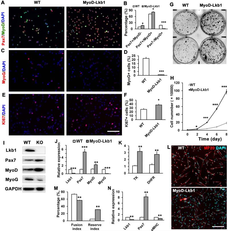
Figure 5.
Lkb1 deletion promotes proliferation but inhibits differentiation of satellite cell derived primary myoblasts. (A-B) Representative images of primary myoblasts from WT and MyoD-Lkb1 mice labeled with Pax7 (red) and MyoD (green), and the percentage of quiescent (Pax7+MyoD-), proliferating (Pax7+MyoD+) and differentiating (Pax7-MyoD+) cells. Nuclei were stained with DAPI (blue). (C-D) Myoblasts stained with differentiation marker MyoG (red) and DAPI; and percentage of MyoG+ cells (D). (E-F) Myoblasts stained with proliferating marker Ki67 (red) and DAPI, the percentage of Ki67+ cells. (G) Colony assay comparing growth of WT and MyoD-Lkb1 myoblasts. (H) Growth curve analysis of the cultured WT and MyoD-Lkb1 myoblasts. (I) Western blot analysis of protein extracts from WT and MyoD-Lkb1 myoblasts. (J-K) mRNA levels of the myogenic (J) and proliferation (K) marker genes in the WT and MyoDLkb1 myoblasts. (L-N) WT and MyoD-Lkb1 myoblasts were differentiated for 3 days. Shown are the myotube morphology (indicated by MF20 staining, Red), fusion index (% nuclei in mytubes), and reserve cell index (% nuclei that are MF20-), and relative mRNA levels of Lkb1, Pax7 and eMHC. Error bars represent SEM, n = 5-7. * P< 0.05, ** P< 0.01, *** P< 0.001. Scale bars: 100 μm.

