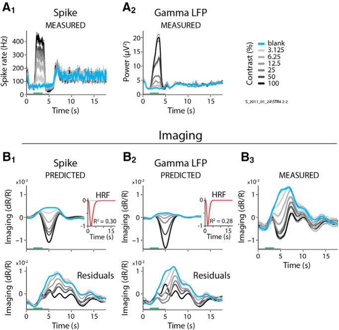
Figure 9.
Full, non-blank-subtracted imaging is poorly fit by spiking and gamma LFP. A1–B3, Single case showing the full (i.e., non-blank subtracted) spiking (A1) and LFP gamma (A2), the corresponding predictions (B1 and B2), and the measured full imaging response (B3; same example as in Fig. 2, following the same conventions). Note the prominent positive signal for the blank and low-contrast stimuli (compare with blank-subtracted measured imaging in Fig. 2B3). Insets depict the optimal HRFs, with fits consistently poorer than for the corresponding blank-subtracted responses (compare Fig. 2B1–B3, insets). Note the large residuals (bottom panels), particularly for low-stimulus contrasts, which are comparable in amplitude to predicted signals.

