Figure 1.
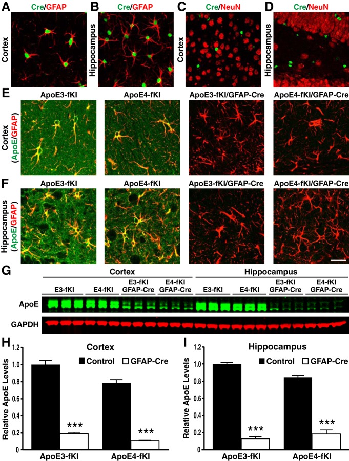
Generation and characterization of apoE-fKI/GFAP-Cre mice. A, B, Representative images of fluorescent immunostaining with anti-Cre recombinase (green) and anti-GFAP (red) in the cortex (A) and hippocampus (B) of apoE-fKI/GFAP-Cre mice. C, D, Neurons immunostained with anti-NeuN (red) did not express Cre-recombinase in the cortex (C) or hippocampus (D) of apoE-fKI/GFAP-Cre mice. E, F, Anti-apoE (green) and anti-GFAP (red) double immunostaining revealed that apoE expression was dramatically decreased in cortical (E) and hippocampal (F) astrocytes in apoE3-fKI/GFAP-Cre and apoE4-fKI/GFAP-Cre mice. G, Representative fluorescent Western blot of apoE (green) and GAPDH (red) in cortical and hippocampal lysates of 17-month-old female mice with different genotypes. H, I, Quantification of apoE protein levels relative to GAPDH in cortical (H) and hippocampal lysates (I) of 17-month-old mice (n = 5/genotype). ApoE levels in apoE3-fKI mice were normalized to 1, and apoE levels in other groups of mice were presented relative to those in apoE3-fKI mice. ***p < 0.001 (t test). Scale bar, 50 μm.

