
Figure 2.
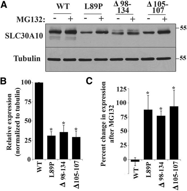
Rapid turnover of SLC30A10 mutants. A, HeLa cells were transfected with indicated FLAG-tagged SLC30A10 constructs. Two days after transfection, cultures were treated with or without 0.5 μm MG132 for 24 h. Cells were then lysed, and FLAG and tubulin were detected by immunoblot. B, Quantification of SLC30A10 levels from A. Expression of SLC30A10 from cultures that were not exposed to MG132 was quantified using NIH ImageJ, and the results were normalized to tubulin expression. Normalized expression of SLC30A10–WT was set to 100 (n = 3 experiments; *p < 0.05 for the difference in expression between each SLC30A10 mutant and WT using one-way ANOVA and Dunnett's post hoc test). C, Quantification of the MG132-induced increase in expression of SLC30A10 mutants from A. SLC30A10 levels after MG132 treatment were quantified using NIH ImageJ and normalized to tubulin. Percentage increase in SLC30A10 expression was then calculated using data in B (n = 3 experiments; *p < 0.05 for the difference between SLC30A10–WT and each mutant by one-way ANOVA and Dunnett's post hoc test. MG132 treatment did not induce a statistically significant change in SLC30A10–WT levels).