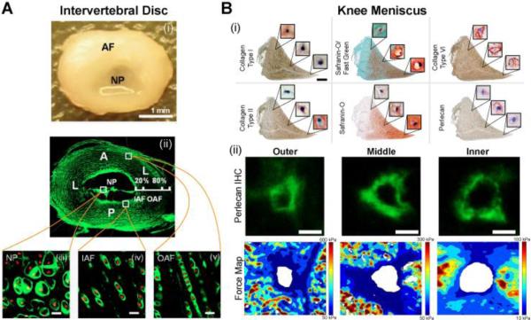
Figure 5. The pericellular matrix in other cartilaginous tissues.
(A) (i) The intervertebral disc is composed of the annulus fibrosis (AF) and nucleus pulposus (NP); (ii) immunofluorescence for type VI collagen in the disc; (iii-v) collagen type VI surrounding cells in the NP, inner AF, and outer AF (scale bar = 20 μm). [Adapted from (Cao et al., 2007), with permission]. (B) (i) Knee meniscus histology and immunohistochemistry showing the variation in ECM and PCM labeling with meniscus region (scale = 0.2 mm, cell view: 20×20 μm); (ii) representative images of perlecan immunolabeling and elastic modulus maps in the outer, middle and inner regions of the knee meniscus [adapted from (Sanchez-Adams et al., 2013), with permission].

