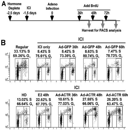
FIG. 3.
Elevation of ACTR promotes cell cycle progression in antiestrogen-treated quiescent cells. (A) Schematics of the time course for treatment of T-47D cells for cell cycle analysis by FACS (see Materials and Methods for details). (B) Two-dimensional FACS distribution of T-47D cells treated as above, grown in regular medium (Regular), or hormone deprived for 60 h (HD), or treated with 10−7 M E2 (E2 48 h), and then labeled with BrdU for 10 h prior to harvesting and staining with propidium iodide. The horizontal axis in the dot plots reflects DNA content, and the vertical axis shows DNA synthesis. The populations of cells in G1 and S phases are indicated. Cells mock infected showed no difference in cell cycle distribution from cells infected with adeno-GFP (data not shown).

