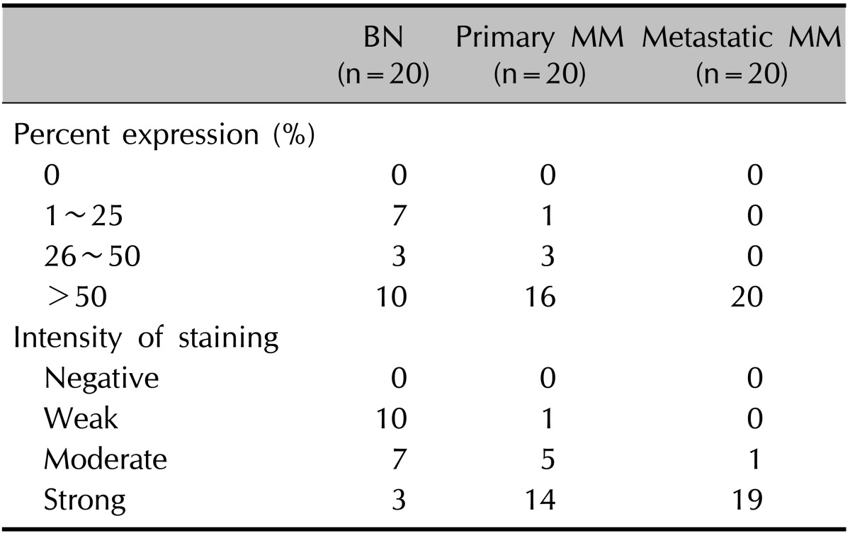
Table 1.
Expression of CacyBP/SIP in MM and BN

After immunohistochemical staining, 3 high-power field images (HPFs, ×200) were randomly selected in each specimen, 100 tumor cells were counted in each field, and the mean percentage of positively stained cells from the 3 HPFs was calculated. Percent expression was graded semiquantitatively as 0%, 1~25%, 26~50% or >50% of the tumor cells were stained. Intensity of staining was graded semiquantitatively as negative, weak, moderate, or strong, respectively. CacyBP: calcyclin-binding protein, SIP: Siah-1 interacting protein, MM: malignant melanoma, BN: benign melanocytic nevus.
