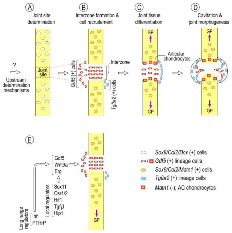
Fig. 2.
Model of limb joint formation and morphogenesis. (A) At early developmental stages, as yet unknown upstream determination mechanisms would identify and prescribe the location of the joints along Sox9/Col2/Dcx-expressing anlagen. (B) Soon after, Gdf5 expression would be activated along with other interzone-specific genes (see E) that would define the initial interzone mesenchymal population within the (Sox9/Col2/Matn1-positive cartilaginous anlagen. This would be accompanied by cell immigration from the flank, and cells located dorsally and ventrally would activate Tgfbr2 expression. (C) Gdf5-positive cells adjacent their respective cartilaginous anlagen -with a Sox9/Col2 history but negative for matrillin-1 expression- would differentiate into articular chondrocytes. (D) Additional differentiation processes and mechanisms such as muscle movement would bring about cavitation and genesis of other joint tissues such as ligaments and other meniscus involving Gdf5- and Tgfbr2-positive and -negative cell progenies. Note that the above distinct spatio-temporal steps -presented here as distinct for illustration purposes- may actually occur more closely and involve overlapping events. Also, the model may not entirely apply to other joints -including intervertebral and temporomandibular joints- that involve additional and/or diverse mechanisms. (E) Schematic summarizing local and long-range regulators that converge to regulate interzone gene expression at early stages of joint formation. Note that this list is not exhaustive.

