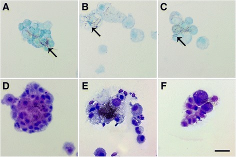
Figure 4.

L-Ag NWs produce frustrated phagocytosis. Panels are Brightfield microscopy images of cells recovered from rat BALF at Days 1 (left), 7 (middle), and 21 (right) after a single instillation (1.0 mg/kg ) of L-Ag NWs (indicated by black arrows). BAL cells were stained using autometallography with a toluidine blue counter-stain (A-C), or Diff Qwik® (D-F). Images from slides stained with autometallography and Diff Qwik® are presented to enable the reader greater identification of Ag NW and cell types, respectively. Scale bar is 25 μm.
