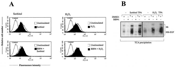
FIG. 4.
Analysis of pro-HB-EGF release in response to stress agents. (A) Flow cytometric analysis of pro-HB-EGF processing. Cos-7 cells were pretreated with BB94 (10 μM) or an equal volume of empty vehicle (dimethyl sulfoxide [DMSO]) for 20 min and stimulated with 0.3 M sorbitol or 200 μM hydrogen peroxide for 30 min. Cells were collected and stained for surface pro-HB-EGF and analyzed by flow cytometry. Control cells were labeled with fluorescein isothiocyanate-conjugated secondary antibody alone. (B) Immunoblot analysis of conditioned media. Cos-7 cells were transiently transfected with pro-HB-EGF cDNA. After serum starvation for 24 h cells were stimulated for 20 min with sorbitol (0.3 M) or hydrogen peroxide (200 μM), and proteins within the supernatant medium were precipitated by TCA precipitation. Precipitated proteins were subjected to Tricine-SDS gel electrophoresis according to the protocol of Schägger and von Jargow and subsequent immunoblot analysis with anti-HB-EGF antibody. TPA stimulation has been included as a positive control. The data shown are representative of three independent experiments.

