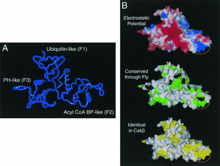
FIG. 3.
Model of the FERM domain of FAK. (A) The backbone of the model of the FERM domain of FAK is shown. This model contains FAK residues 60 to 349. The ubiquitin-like subdomain (F1), acyl-CoA binding protein (BP)-like subdomain (F2) and PH/PTB/EVH-like subdomain (F3) are indicated. (B) The surface of the model of the FERM domain is shown. In the top panel, electrostatic potential is indicated colorimetrically, with red indicating negative potential and blue indicating positive potential. In the middle panel, the surface of the FERM domain is shown with residues that are identical from Drosophila to human (green) and highly conserved residues (black) indicated. In the bottom panel, the surface of the FERM domain is shown with residues that are identical between FAK and CAKβ in yellow. The circled region indicates a highly conserved basic patch at the tip of the F2 subdomain.

