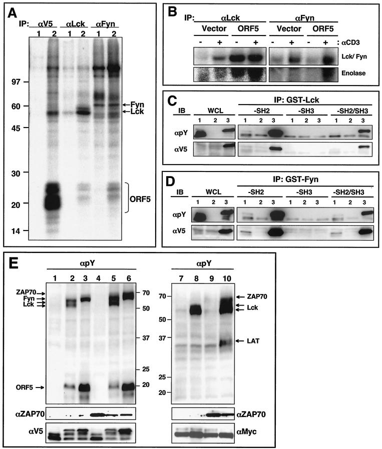
FIG. 5.
Interaction of ORF5 with Src family kinases. (A) In vitro immune complex kinase reactions. Jurkat-vector (lanes 1) and Jurkat-ORF5 (lanes 2) cell lysates were immunoprecipitated (IP) with anti-V5 (αV5), anti-Lck, and anti-Fyn antibodies. Subsequently, immunoprecipitates were subjected to an in vitro immune complex kinase assay with [γ-32P]ATP. Arrows indicate Fyn, Lck, and ORF5 proteins. Molecular weight markers (in thousands) are indicated at the left. (B) In vitro Lck and Fyn kinase reactions. Jurkat-vector and Jurkat-ORF5 cells were unstimulated (−) or stimulated (+) with an anti-CD3 antibody for 3 min, and cell lysates were then immunoprecipitated with anti-Lck and anti-Fyn antibodies. Subsequently, immunoprecipitates were subjected to an in vitro immune complex kinase assay with [γ-32P]ATP and 5 μg of enolase. (C) In vitro interaction of tyrosine-phosphorylated ORF5 with the SH2 domain of Lck kinase. 293T cells were transfected with the V5-tagged ORF5 expression vector (lanes 1), Myc-tagged Lck expression vector (lanes 2), or V5-tagged ORF5 and Myc-tagged Lck expression vector (lanes 3). Whole-cell lysates (WCL) were used for immunoblotting (IB) with anti-V5 or antiphosphotyrosine (αpY) antibody to show ORF5 expression and its tyrosine phosphorylation. The same WCL were then used for binding assay with Sepharose bead-conjugated Lck-SH2, Lck-SH3, or Lck-SH2/SH3 followed by immunoblotting with either an anti-V5 or antiphosphotyrosine antibody. (D) In vitro interaction of tyrosine-phosphorylated ORF5 with the SH2 domain of Fyn kinase. Samples and procedures were as described for panel C, except for the use of Fyn in place of Lck kinase. (E) Lck and Fyn but not ZAP70 phosphorylate ORF5. 293T cells were transfected with the V5-tagged ORF5, Myc-tagged LAT, Lck, Fyn, and ZAP70 expression vector in various combinations. At 48 h posttransfection, cell lysates were used for immunoblotting with an antiphosphotyrosine antibody. The bottom panels show the expression levels of ZAP70, ORF5, and LAT. Arrows indicate Lck, Fyn, ZAP70, LAT, and ORF5. Lane 1, ORF5; lane 2, Lck plus ORF5; lane 3, Fyn plus ORF5; lane 4, ZAP70 plus ORF5; lane 5, Lck plus ZAP70 plus ORF5; lane 6, Fyn plus ZAP70 plus ORF5; lane 7, LAT; lane 8, Lck plus LAT; lane 9, ZAP70 plus LAT; lane 10, Lck plus ZAP70 plus LAT. Numbers on the left are molecular weights in thousands.

