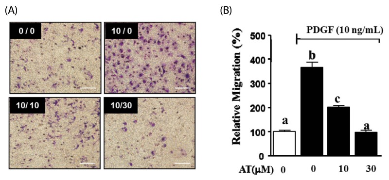
Fig. 3.
Effects of AT on migration in PDGF-BB-stimulated VSMC. VSMC were treated with AT (10 µM and 30 µM) and PDGF-BB (10 ng/mL) for 90 min. (A) Migrating VSMC on membranes, the spots are Diff quick-stained cells. The number at the top right corner of each image is the concentration of PDGF-BB/concentration of AT. Magnification, × 200. Bar = 500 µm. (B) Migrated cell counts indicated relative to the untreated control (%). Results are presented as mean ± standard error. Values with the same superscript letter are not significantly different by Duncan's multiple range test (P < 0.05).

