
Figure 7.
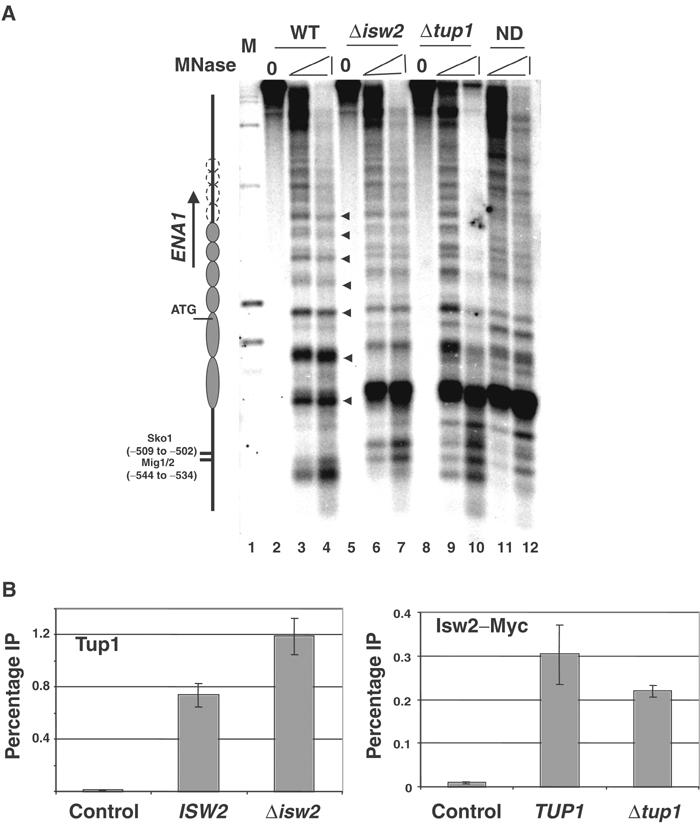
Collaboration of Ssn6–Tup1 and ISW2 at the ENA1 locus. (A) MNase mapping of the ENA1 promoter detected by indirect end labeling. Filled triangles represent the regularly spaced hypersensitive sites in the wild-type chromatin, which are interpreted as internucleosomal sites. The open circles extending beyond nucleosome 6 (left of panel) indicate the possibility that nucleosome positioning continues, but could not be resolved by this gel. (B) ChIP assay for Tup1 and Isw2-Myc crosslinking to the ENA1 URS. Tup1 and Myc antibody were used, with the preimmune or IPs from an untagged strain used as negative controls, respectively. The PCR fragment corresponding to −632 to −316 relative to the translation start site flanks the Sko1 and Mig1/2 binding sites.